Humans breathe in a mix of air containing roughly 21% oxygen—yet, the oxygen levels in our individual cells vary considerably, depending upon their location and function and our body’s overall state.
Cells also self-regulate their oxygen levels to suit their activities, consuming more as needed. It’s a dynamic environment that adapts to demands and conditions. However, when trying to study cell behavior in the lab, researchers have historically relied upon experimental cell culture setups with decidedly unnatural oxygen environments.
Most research labs run their cultures at the ambient oxygen level of 21%; some use gas regulators to strictly control the level, which still doesn’t allow for the homeostatic processes that play out in the body.
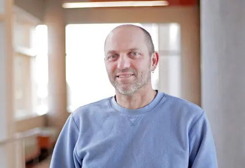
 David Beebe, the John D. MacArthur Professor and Claude Bernard Professor of biomedical engineering, has made fundamental contributions to the field of microfluidics.
Those approaches—standard lab protocols—are akin to studying exercise science by asking humans to work out at dangerously high or low oxygen levels or in a rigidly set oxygen tent, says David Beebe, a professor in the Departments of Biomedical Engineering and Pathology and Laboratory Medicine at the University of Wisconsin-Madison.
Beebe and some of his lab members believe they’ve found a better way to mimic the body’s natural oxygen microenvironment by entrusting the cells themselves to regulate the oxygen level in microscale cell cultures.
In a paper appearing in the April 5 issue of Advanced Science, Beebe, scientist Chao Li and collaborators from UW-Madison and beyond present their solution: an under-oil, microfluidic cell culture system that allows cells to autonomously regulate their oxygen microenvironment, which researchers can dynamically monitor.
“Nobody’s ever done this in a way that is so simple. It’s really simple for anybody to use this,” says Beebe, who specializes in developing straightforward technology for cell culture applications. “We will likely see different biology now, and the argument would be that it will be more relevant biology.”
Building upon its previous work with under-oil microfluidic systems, the Beebe Lab’s method uses silicone oil to achieve the “just right” level of oxygen permeability (hence the Goldilocks reference in the paper’s title), along with an optical sensor and a dye to monitor oxygen concentration at both the cell layer and from within the cultured cells.
“It’s super easy to just pipette in different amounts of oil to have different thicknesses and find that sweet spot where the cells are able to control that environment,” says Beebe, noting the method doesn’t require researchers to build or acquire a new device or system.
The researchers tested their setup across a range of mammalian and fungal cells, culminating with a successful coculture of intestinal cells and one type of bacteria commonly found in the human gut. That’s an especially useful demonstration, Beebe notes, given that the intestinal cells require oxygen while the bacteria are intolerant of it.
“It proved that by simply putting the right things together in the culture system, we can mimic the in vivo environment without using any complicated external equipment,” says Li, the first author on the paper.
Ophelia Venturelli, an assistant professor of biochemistry who led genomic editing and analysis elements of the project, says the group’s simple coculture approach could be especially useful in further probing interactions between host cells and microbial communities and uncovering the molecular mechanisms at play.
Li can trace the project back to his early days in the Beebe Lab in 2017, when he questioned the oxygen level setting on an incubator. He says discovering a range of oxygen levels within different cell types and oil overlays was a fortuitous observation during earlier development of the group’s under-oil microfluidic system.
Nearly five years after first discussing the prospect of allowing cultured cells to self-regulate their oxygen levels with Beebe, Li found himself explaining the basic concepts to his 6-year-old daughter, Annie. He and his coauthors needed an image to submit to the journal, and Annie was a budding artist who was game for the challenge.
She molded her Play-Doh into a replica Goldilocks, along with three sets of oxygen levels—too much, too little and just right. Advanced Science chose it as a full-page image to pair with the article.
David Beebe is the Claude Bernard Professor of Biomedical Engineering and the John D. MacArthur Professor in the Department of Pathology and Laboratory Medicine in the UW-Madison School of Medicine and Public Health. Glenn Walker (PhDBME ’02), a Beebe Lab alumnus and associate professor at the University of Mississippi, provided computational modeling to support the research. Ophelia Venturelli is an affiliate with the Department of Chemical and Biological Engineering. Researchers from the UW-Madison School of Pharmacy’s Mass Spectrometry Facility also assisted with analysis.