Complex human joints are a challenge to visualize. Textbook figures may be helpful, but they only offer a static, two-dimensional view. Meanwhile, real-life joints in actual humans are difficult to gain access to, unless you’re an orthopedic surgeon—not to mention that they’re inevitably obscured by a mess of blood and body tissue.
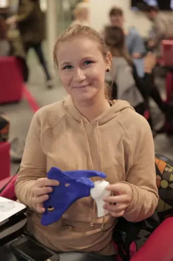 Students in the course develop computer models of their joint replacements and then print them with a 3D printer at the new makerspace. Photo: Renee Meiller.
So, what is an engineering student interested in orthopedic joints and implants to do? For starters, they could sign up for Mechanical Engineering Associate Professor Heidi Ploeg’s course, Design of Orthopedic Implants, which makes innovative use of UW-Madison’s most cutting-edge learning technology in the College of Engineering’s new makerspace, the Grainger Engineering Design Innovation Laboratory.
Students in Ploeg’s course for seniors and graduate students use the makerspace’s enormous interactive touchscreen and virtual reality headset to study bone joint anatomy. They use the technology to study computed tomography (CT) scan data from the National Library of Medicine, which provides the basis for the total joint replacements they’ll develop using computer modeling. The 10 teams of four students each can then use the visualization tools to examine their implant models before they finally print them, also at the makerspace, with a 3D printer.
Ploeg, who has taught the course in previous semesters, says the new makerspace technology has opened up new avenues for assisting engineering students in their learning. The students learn about joint anatomy through textbooks and a visit to the UW Anatomy Lab, hosted by resident anatomy expert Ed Bersu.
“This semester we were able to provide an extra learning experience that we weren’t able to before,” Ploeg says, referring to the visualization lab and 3D printer. “The visualization lab provides a nice environment for the students to collaborate and share their ideas as they work on models.”
Within 75 minutes of providing students with the CT scan data for the human joints they would be tasked with replacing, Ploeg says each of the 10 teams had a computer model ready to share. “I think it helps that we’re in this cool room where we can interact with the models with shared screens including this huge touchscreen,” Ploeg says.
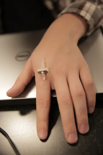 A student team is working on developing a knuckle joint replacement with titanium sleeves at either end to make implant replacement quick and simple. Photo: Renee Meiller.
The ability to 3D print their models was the cherry on top of an already fun learning experience, Ploeg adds. “Even when you’re able to see and interact with a model on a screen, it’s a totally different experience to be able to interact with the model physically,” Ploeg says. “You see, and feel, more details and can understand it better. It’s a whole different interaction.”
Molly Scott, a senior biomedical engineering student, is a member of a team in Ploeg’s class working on modifications to the standard knuckle joint replacement. She and her team want to develop an implant with titanium sleeves at either end that are mounted onto the bones and make implant replacement quick and simple.
“We’re focusing on a younger patient demographic,” Scott says. “Younger people who receive a joint replacement like this almost certainly will have to have it replaced at some point because the silicone implant will eventually fail.”
The idea behind the titanium sleeves is to make future replacement operations as non-invasive as possible.
Scott says the experience using the makerspace technology has been an enjoyable one.
“I’ve been like a kid in a candy shop with everything here in the makerspace,” Scott says. “Nothing is ever engineered in a vacuum, so it’s a lot cooler to see the implants we’re developing physically and be able to study the joints with the vis lab screen and the 3D models. We actually get to put the information we’re learning into action and test it.”
Scott says she’ll likely pursue a career in the field, and this experience helped confirm her enthusiasm for it.
“In the future, we’re moving away from one-size-fits-all joint replacements; rather, patients will come to expect joints that are tailored to them individually,” Scott says.
And the technology—CT scan data, high-powered visualization tools and methods to cheaply fabricate 3D models—that will make that personalization possible is the same type of technology Ploeg’s class is using right now.