When pathologists examine tissue samples for signs of disease, they rely on their trained eyes to spot changes in specific biomarkers that indicate abnormalities.
Before those alterations become visible on chemically stained samples, though, disease-related manipulations occur at the molecular level.
A new imaging system featuring a novel application of engineered plasmonic metasurfaces, devised by University of Wisconsin-Madison biomedical engineers, can chemically map tissues, providing a more detailed, quantitative and representative picture. In addition to enhancing clinical histopathology capabilities, it could aid toxicology studies and reveal new clues into the origins and early markers of various diseases.
The researchers, led by Filiz Yesilköy, an assistant professor of biomedical engineering, demonstrate their approach by imaging brain tissue in a paper in the journal Advanced Materials. By pairing advanced microscopy tools with their unique nanofabricated metasurface to boost mid-infrared imaging capabilities, Yesilkoy and PhD student Samir Rosas can detect a wider range of biomolecules while overcoming any bias caused by the thickness or heterogeneity of a given tissue sample.
“We are eliminating the correlation to the physical bulk tissue properties. We are just confining and quantitatively analyzing the tissue material in the nanoscale,” says Yesilkoy, whose lab studies photonics and develops nanotechnology for imaging and sensing tools. “There are lots of diseases where, first of all, we don’t have the biomarkers specific to stain the tissue against. So in that case, we need to understand the overall chemical composition of the tissue. And with today’s artificial intelligence and machine learning algorithms, if we have this vast amount of chemical information that is resolved spatially, then we can benefit from this data science analytics.”
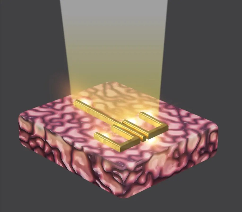 Rendering of the plasmonic metasurface. Image courtesy Yesilkoy lab.
Their method harnesses the unique properties of the plasmonic metasurface Rosas developed in the College of Engineering’s Nanoscale Fabrication Center over the course of two years. The collective effect of almost three million gold resonators adorning the metasurface allows it to cover a wider range of frequencies, and thus, biomolecules, when tissue is placed atop it.
“The plasmonic metasurface has several remarkable features,” says Rosas, citing its abilities to cover the desired spectral range, extract molecular information from ultra-thin tissue, and yield information from the same diffraction-limited spot. “Furthermore, our method is completely label-free.”
Yesilkoy and Rosas can even image tissue regions that wouldn’t otherwise be visible, as well as detect the specific structural alignment of proteins. The latter capability could prove particularly useful for studying neurodegenerative diseases, such as Alzheimer’s and Parkinson’s, which are characterized by a buildup of misfolded proteins.
“We see a lot of prospects for studying neurodegenerative diseases, because it has that niche where the protein structure can be studied using this method, contrary to other brain imaging and tissue imaging methods,” says Yesilkoy.
She and Rosas plan to continue working with collaborator Xinyu Zhao, a professor of neuroscience at UW-Madison whose lab provided tissue samples for the work, to examine brain tissue from mice that have been genetically modified to carry autism. The researchers will use their new technique to create chemical maps of the brains of mice at different ages and identify physiological changes.
“When we don’t know what we’re looking for,” says Yesilkoy, “looking for everything and just feeding that into the data processing pipeline can actually illuminate it.”
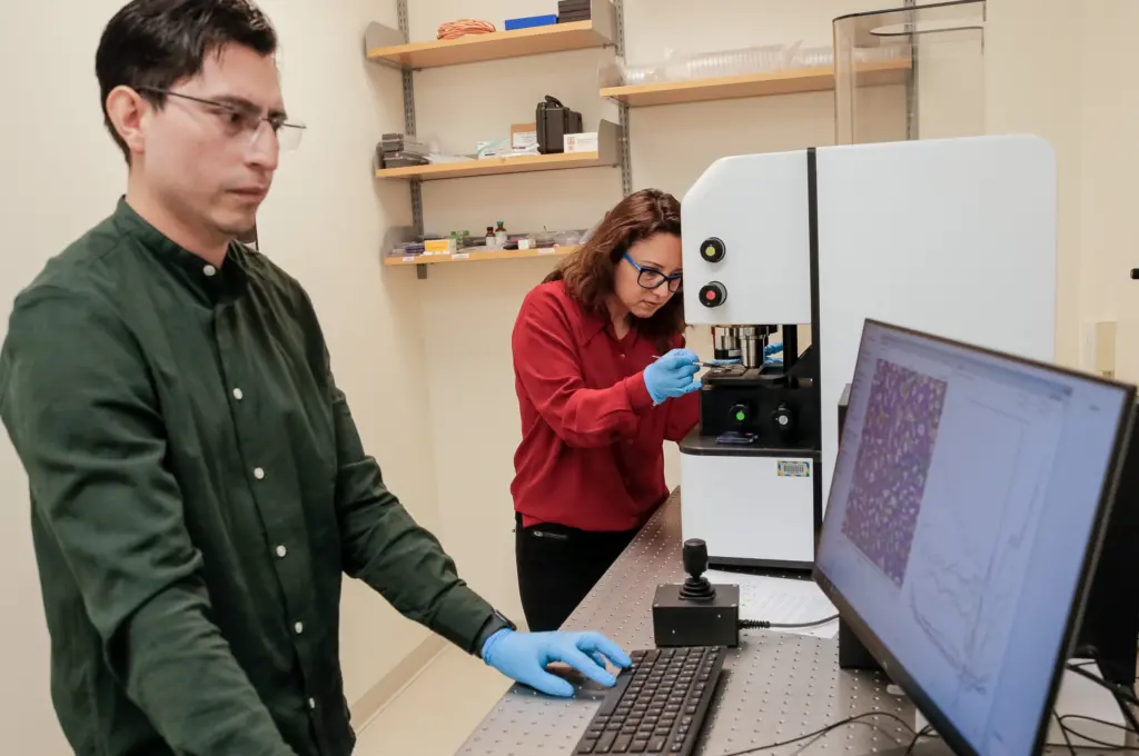 PhD student Samir Rosas and Assistant Professor Filiz Yesilkoy. Photo by: Tom Ziemer.
Other UW-Madison authors on the paper include: Kevin Eliceiri, an associate professor of biomedical engineering and medical physics and the Retina Research Foundation Walter H. Helmerich Research Chair at the McPherson Eye Research Institute; Mikhail Kats, Jack St. Clair Kilby associate professor of electrical and computer engineering and H. I. Romnes Faculty Fellow; Hongyan Mei, a PhD student in electrical and computer engineering; Edward Chang, an undergraduate neurobiology major; and Keegan Schoeller, a recent graduate in biology.
Top photo caption: Assistant Professor Filiz Yesilkoy and PhD student Samir Rosas show one of their lab’s engineered plasmatic metasurfaces mounted with brain tissue. Photo by: Tom Ziemer.