By creating a miniscule device that translates brain activity into radiowaves, University of Wisconsin-Madison neuroengineers have taken a key step toward enabling injectable sensors for wireless, localized recording in the brain.
The work, detailed in the May 2023 issue of the journal Sensors and Actuators B: Chemical, is essentially a proof-of-concept for using minimally invasive sensors that wirelessly harvest power and transmit data to external imaging hardware operating on radio frequency, such as magnetic resonance imaging (MRI) scanners.
“Right now, in order to get brain recordings of any description, you’ve got to have either a wired probe or a wireless probe with bulky hardware on top of your head,” says Suyash Bhatt, a PhD student in biomedical engineering and co-first author on the paper along with undergraduate Emily Masterson. “In our case, you can actually inject this into the brain, look at what’s going on in MRI. The MRI provides the power, and you don’t even need anything else in terms of circuitry. So that’s a pretty big step forward.”
While the sensor is broadly applicable, it would be particularly useful for diagnosing and monitoring neurological conditions, including epilepsy, Parkinson’s disease and traumatic brain injury.
The research is part of Biomedical Engineering Assistant Professor Aviad Hai’s quest to create dramatically less invasive technologies that yield more detailed, granular and comprehensive data from across the brain.
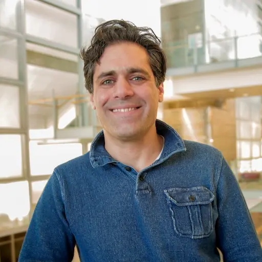
 Assistant Professor Aviad Hai
The Hai lab is creating milliscale sensors that consist of an inductor and capacitor—which form a resonator, essentially acting as an antenna that can absorb electromagnetic energy from an outside transmitter—along with an ion-sensitive field effect transistor. That transistor, powered by the resonator, detects the rapid flow of ions in the brain, which corresponds to neural activity.
To validate their prototype, the researchers surgically implanted it in the brains of rats and then electrically stimulated one of their hind paws, causing a neural response.
“Nothing like this has been shown in the living brain,” says Hai, whose lab has previously published work modeling the use of nanoparticles in conjunction with magnetic particle imaging and nano antennas that can form connections with individual neurons.
Bhatt has already fabricated a microscale version of the device using tools in the College of Engineering’s Nanoscale Fabrication Center. At less than a square millimeter in total area, the sensors are small enough to fit in a 16-gauge needle.
“These are basically just the equivalent of dust,” says Bhatt, who’s planning to pursue medical school after completing his PhD in 2024.
The group also can track specific neurotransmitters—dopamine, for example—by coating the transistor with different chemicals.
Hai hopes to eventually translate the research to clinical use. “There’s no reason not to. We’re using purely biologically compatible materials that have been used for other technologies that are FDA approved,” he says.
For Masterson, claiming a co-first author credit on a paper—a rarity for an undergraduate—is a satisfying reward to more than three years in the lab. As a first-year student, she sought out the research opportunity, and subsequently earned a Sophomore Research Fellowship and a Hilldale Research Fellowship to support her work. When the COVID-19 pandemic shuttered campus in 2020, Hai shipped kits for making microelectrodes to Masterson’s house in North St. Paul, Minnesota, where she built them in her parents’ basement while Zooming with Hai in Madison and fellow undergraduate researcher Jenna Eizadi in California.
After graduating in May 2023, Masterson will start a full-time position at Medical Murray, a medical device contractor.
“My research experience has given me the opportunity to accomplish something meaningful and advance the field of neuroscience,” says Masterson, who presented with Bhatt at the Society for Neuroscience’s annual meeting in fall 2022. “Moving forward, I hope to leverage my experience in research to add a different perspective to any conversation.”
Aviad Hai is part of the Wisconsin Institute for Translational Neuroengineering and an affiliate of the Department of Electrical and Computer Engineering.
All authors on the paper are members of the Hai lab. They also include PhD students Tianxiang Zhu, Adam Vareberg, Jack Phillips and Ilhan Bok; UW-Madison graduates Jenna Eizadi (BSBME and computer science ’21), Judy George (MSBME ’21), Nesya Graupe (BSBME ’23),Matthew Dwyer (MSEE ’11, PhDEE ’17); and scientist Alireza Ashtiani (MSEE ’12, PhDEE ’16).
The research was supported by grants from the National Institute of Neurological Disorders and Stroke and the Office of the Director’s Common Fund at the National Institutes of Health (grant DP2NS122605); the National Institute of Biomedical Imaging and Bioengineering (grant K01EB027184); the Office of Naval Research (grants N00014-23-1-2006 and N00014-22-1-2371); and the Wisconsin Alumni Research Foundation (WARF).
Top photo caption: PhD student Suyash Bhatt and undergraduate Emily Masterson are using an MRI machine to validate the Hai lab’s minimally invasive sensors. Photo: Tom Ziemer.